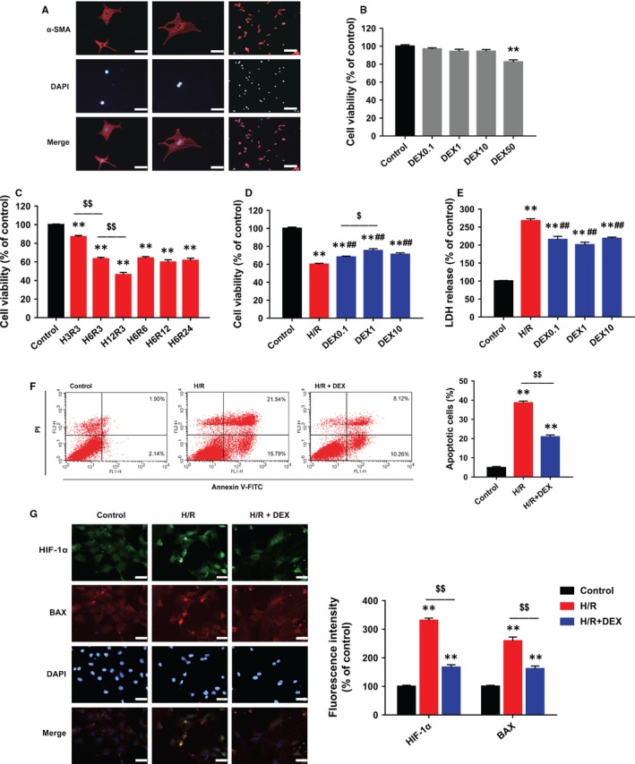
Figure 2.

DEX post‐treatment alleviated H/R‐induced injury, inhibited apoptosis and reduced HIF‐1α and BAX expression in neonatal rat cardiomyocytes. A, Immunostaining of α‐SMA showing purified cardiomyocytes. Scale bars: 50 (left and middle rows) and 200 µm (right row). B, CCK‐8 assay showing that DEX 0.1‐10 µM had no cytotoxicity. C, CCK‐8 assay showing significant decreases in cell viability after different durations of H/R (eg H3R3 indicates 3 hours of hypoxia followed by 3‐hours of reoxygenation). D, CCK‐8 assay showing the effects of DEX post‐treatment on cell viability. E, LDH assay showing the effects of DEX on cytotoxicity. F, Flow cytometric assessment of annexin V‐FITC/PI staining showing that DEX reduced apoptotic cells. G, Immunofluorescence showing that DEX inhibited the expression of HIF‐1α (green) and BAX (red). Scale bar: 30 µm. n = 6. **P < .01 vs control; ##P < .01 vs H/R; $P < .05, $$P < .01 for the comparisons shown
