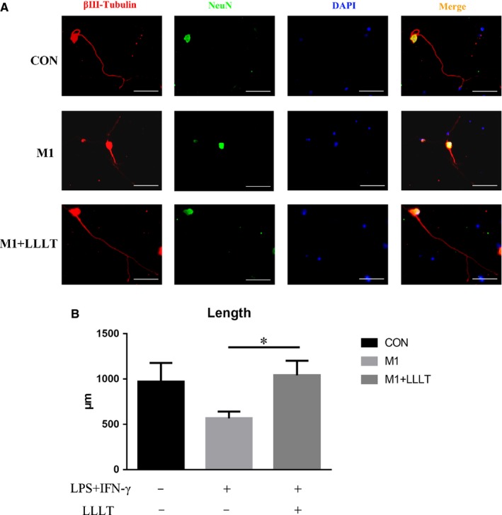
Figure 4.

A, Forty‐eight hours after irradiation, the supernatants were individually collected and added to DRG neurons; after 48 h, immunofluorescence staining was used to assess axonal length with βIII‐tubulin as an axonal marker and NeuN as a neuronal body marker. B, The M1+LLLT group showed significantly improved neuronal growth compared with the M1 group. Bar = 200 μm. *P < .05 compared with the M1 group
