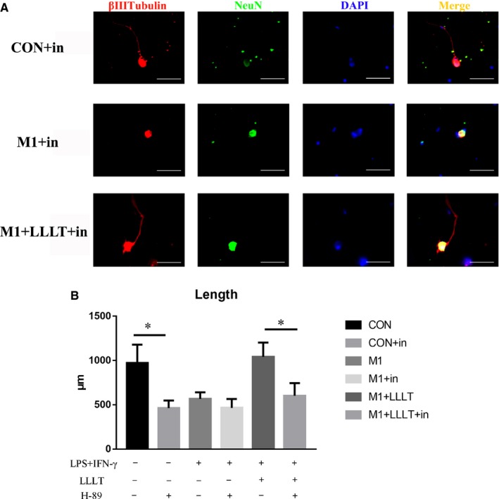
Figure 9.

A, Immunofluorescence staining was used to analyse the length of axons, with βIII‐tubulin as an axonal marker and NeuN as a neuronal body marker. B, The axon length of each group was compared according to inhibitor presence. *P < .05, **P < .01, ***P < .001
