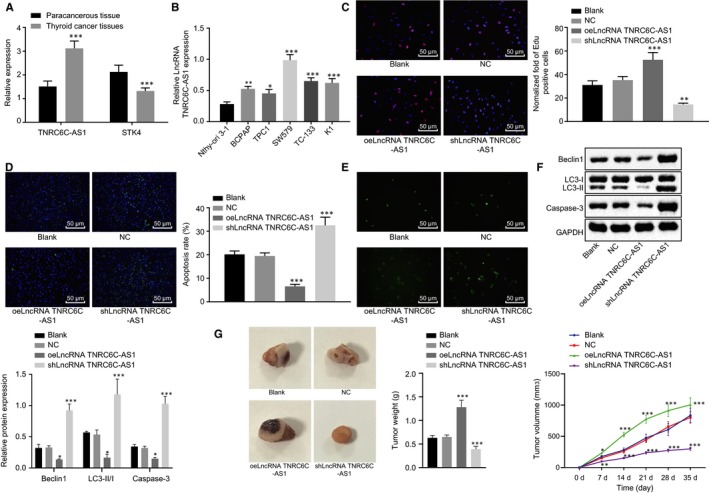
Figure 2.

LncRNA TNRC6C‐AS1 silencing inhibits proliferation and promotes apoptosis and autophagy of TC cells in vivo. A, The expression of lncRNA TNRC6C‐AS1 and STK4 in 54 cases of TC tissues and adjacent tissues were analysed. ***P < .001 vs the adjacent tissues, (n = 54), the comparison between the two groups was analysed by t test (mean ± standard deviation). B, The expression of lncRNA TNRC6C‐AS1 in 5 TC cell lines was determined by RT‐qPCR, with normal human thyroid cell Nthy‐ori 3‐1 as a reference. *P < .05, **P < .01, ***P < .001 vs Nthy‐ori 3‐1. C, EdU assay was used to detect the proliferation ability of SW579 cells transfected with oelncRNA TNRC6C‐AS1 and shlncRNA TNRC6C‐AS1, respectively. D, TUNEL method was used to detect the apoptosis rate of SW579 cells transfected with oelncRNA TNRC6C‐AS1 and shlncRNA TNRC6C‐AS1, respectively. E, MDC method was used to detect the number of autophagic vesicles in SW579 cells transfected with oelncRNA TNRC6C‐AS1 and shlncRNA TNRC6C‐AS1, respectively, compared with the NC group. F, Western blot analysis was performed to assess the expression of apoptosis (Casepase‐3) and autophagy‐related (Beclin1 and LC3‐II/I) factors in SW579 cells transfected with oelncRNA TNRC6C‐AS1 and shlncRNA TNRC6C‐AS1, respectively, normalized to GAPDH. G, The representative pictures, weight and volume of tumours in nude mice (n = 14). *P < .05, **P < .01, ***P < .001 vs the NC group. The comparison among multiple groups was analysed by one‐way or repeated measures ANOVA (mean ± standard deviation). Values were obtained from 3 independent experiments in triplicate
