Figure 1.

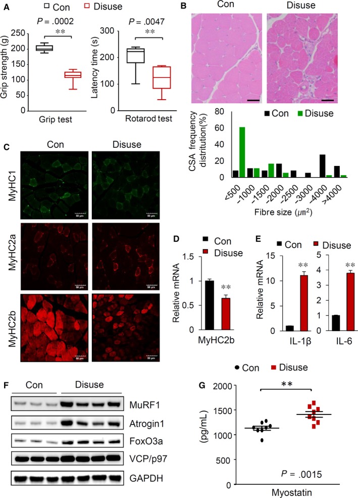
Muscle disuse generated by hindlimb immobilization in mice induces muscle atrophy. A, Grip strength measurements (left) and rotarod test results (right) of the control and hindlimb‐immobilized mice (n = 8). B, A reduction in muscle mass induced by hindlimb immobilization. H&E staining of GA muscle sections (top). Scale bars: 50 μm. Distribution of GA muscle fibres in cross‐sectional areas (bottom). C, Muscle sections prepared from GA muscles of the control and hindlimb‐immobilized mice were visualized with immunostaining against MyHC I, MyHC IIa and MyHC IIb. Scale bars: 50 μm. D, Relative mRNA levels of MyHC2B in the GA muscle (n = 8). E, Changes in the mRNA levels of IL‐1β and IL‐6 in the GA muscle (n = 8). F, Alterations in proteins related to muscle atrophy. Immunoblot analysis of the indicated proteins in the GA muscle. G, Serum myostatin levels in the control and hindlimb‐immobilized mice (n = 8). Serum myostatin levels (n = 8/group) were quantified by using a GDF8/myostatin Quantikine ELISA kit (R&D Systems) according to the manufacturer's instructions. All results are representative of more than three independent experiments. The data represent the mean ± SEM. P values < .05 obtained from Student's t tests were considered significant
