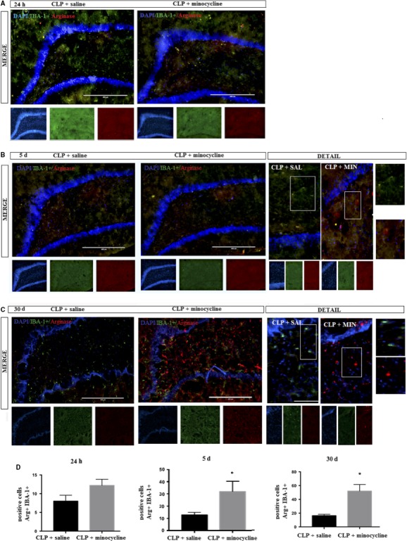
Figure 6.

DAPI (nuclei—blue); arginase (M2 marker—red); IBA‐1+ (microglial marker—green) was determined by immunofluorescence, 24 h, 5 and 30 d after sepsis in the hippocampus of animals treated with or without minocycline. A, 24 h; B, 5 d; C, 30 days; D, corresponding graphs. Image details are presented. Double positive cells (CD11b + IBA‐1+) were counted in both groups. t Test for independent samples. P = .74; P = .03
