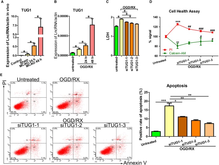
Figure 1.

TUG1 was up‐regulated in MA‐C cells cultured under OGD/R conditions. A, QRT‐PCR showing TUG1 levels in MA‐C cells at 24 or 48 h post‐reoxygenation. B, TUG1 levels were measured using qRT‐PCR at 24 h/48 h post‐reperfusion after MCAO was established. C, LDH leakage was measured using LDH assays after TUG1 knockdown under OGD‐R conditions (6‐h OGD and 24‐h reoxygenation). D, Cell health was measured using cell health assays after TUG1 knockdown under OGD‐R conditions (6‐h OGD and 24‐h reoxygenation). E, Flow cytometry assays were performed to show the cell apoptosis after transfection with short interfering RNAs siTUG1‐1, siTUG1‐2 and siTUG1‐3 after 6‐h OGD/24‐h reoxygenation treatments. MCAO, middle carotid artery occlusion; OGD/R, oxygen‐glucose deprivation and reperfusion; qRT‐PCR, quantitative real‐time reverse‐transcription polymerase chain reaction; TUG1, taurine‐up‐regulated gene 1
