Figure 5.

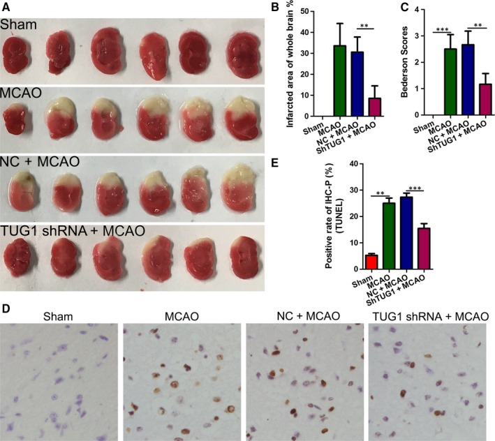
TUG1 shRNA attenuated the infarction area and decreased cell apoptosis in I/R mouse brains. A‐B, Representative coronal sections of TTC staining after MCAO. The relative infarct area percentage was evaluated by observing the unstained infarcted tissue zone (white) and the stained normal tissue zone (red). C, Bederson scores of mice treated with or without TUG1 shRNA after MCAO. D‐E, Cell apoptosis was examined using a TdT‐mediated biotin‐16‐dUTP nick‐end labelling (TUNEL) assay (magnification, ×400; **P < .01, ***P < .001 vs. sham). MCAO, middle carotid artery occlusion; shRNA, short hairpin RNA; TTC, 2,3,5‐Triphenyltetrazolium chloride; TUG1, taurine‐up‐regulated gene 1
