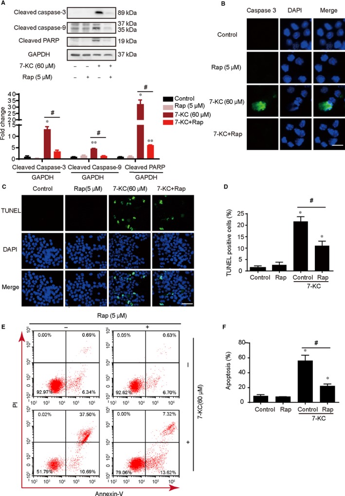
Figure 3.

Enhancement of autophagy by rapamycin attenuated cells apoptosis induced by 7‐KC in RAW264.7 cells. RAW264.7 cells were treated with Rap (5 μmol/L) for 60 min and then exposed to 7‐KC (60 μmol/L) for additional 24 h. A, Western blot analysis of cleaved caspase‐3, cleaved caspase‐9 and cleaved PARP expression in the whole cells. B, Cells were stained by cleaved caspase‐3 (green) and DAPI (blue) and analysed by fluorescence microscopy, scale bar = 15 μm. C, D, Representative images of TUNEL staining of macrophages showed the apoptotic cells (apoptotic cells stained in green and nucleus stained in blue with DAPI). The number of TUNEL‐positive cells was measured and quantitated. Scale bar = 50 μm. E, F, RAW 264.7 cells were cotreated with 60 μmol/L 7‐KC and 5 μmol/L Rap for 24 h compared with control or 7‐KC and Rap treatment alone, and apoptosis was examined by using annexin V with PI staining with flow cytometry. Data were presented as mean ± SEM of at least three independent experiments. *P < .05 vs control, ** P < .01 vs control, #P < .05 vs 7‐KC group
