
Figure 5.
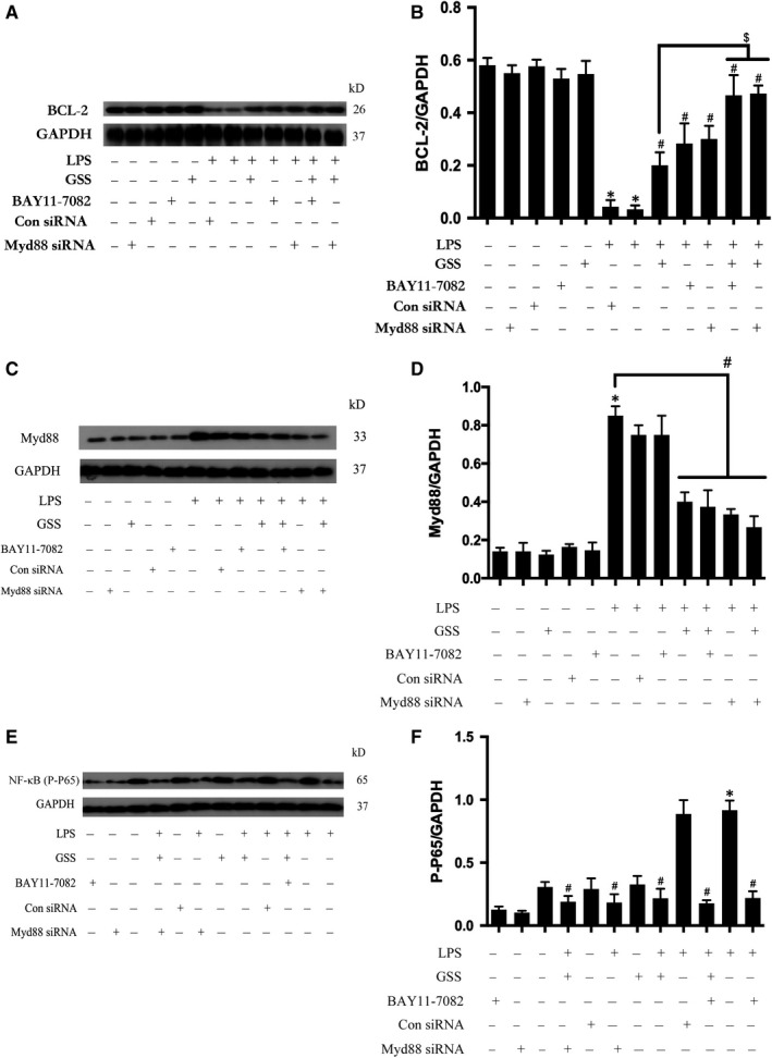
GSS inhibits LPS‐induced EC apoptosis via the Myd88/NF‐κB/BCL‐2 signalling pathway. A, After transfection with Myd88 siRNA for 48 h, ECs were treated with GSS for another 2 h prior to the stimulation with LPS for 24 h, and the expression of BCL‐2 was determined by Western blot. Similarly, ECs were pre‐treated with BAY11‐7082 and GSS for 2 h and then incubated with LPS for 24 h, and BCL‐2 expression was measured by immunoblotting. B, The Western blotting results are presented as a histogram showing the band intensity values. C, After transfection with Myd88 siRNA for 48 h, ECs were treated with GSS for another 2 h prior to the stimulation with LPS for 24 h, and the expression of Myd88 was determined by Western blot. Similarly, ECs were pre‐treated with BAY11‐7082 and GSS for 2 h and then incubated with LPS for 24 h, and the Myd88 expression was checked by immunoblotting. D, The Western blotting results are presented as a histogram showing the band intensity values. E, After transfection with Myd88 siRNA for 48 h, ECs were treated with GSS for another 2 h prior to the stimulation of LPS for 24 h, and the activation of NF‐κB was determined by Western blot. Similarly, ECs were pre‐treated with BAY11‐7082 and GSS for 2 h and then incubated with LPS for 24 h, and the activation of NF‐κB was checked by immunoblotting. F, The Western blotting results are presented as a histogram showing the band intensity values. *P < .05 vs the negative control. # P < .05 vs the corresponding LPS treatment group. $ P < .05 vs the corresponding GSS treatment group