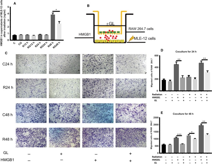
Figure 6.

GL blocks the chemotaxis of HMGB1 in vitro. A, HMGB1 in supernatants from MLE‐12 cells after irradiation at different times by ELISA. HMGB1 was increased only at 48 h after irradiation. B, Scheme of the transwell assay. MLE‐12 cells were seeded in the lower compartment and underwent irradiation, while RAW 264.7 cells were seeded in the upper chamber and co‐cultured for 24 and 48 h, respectively. C, Representative images of migrated cells stained with crystal violet after co‐culturing for different times. 100× D,E, Result of migrated cell counts after co‐culturing for 24 and 48 h. (*P < .5, **P < .01, ***P < .001)
