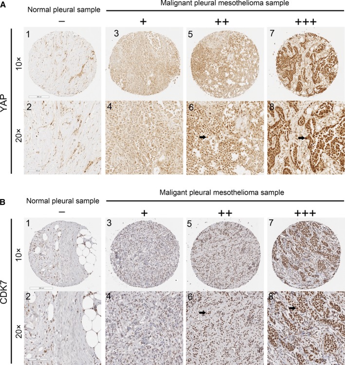
Figure 1.

Immunohistochemistry of CDK7 and YAP in human malignant pleural mesothelioma (MPM) Representative image showing expression of YAP protein (A) and CDK7 protein (B) in human MPM tissues and normal pleural tissues analysed by immunohistochemistry. (A: 1‐2) and (B: 1‐2) are normal pleural tissues. (A: 3‐8) and (B: 3‐8) are MPM tissues. (A: 4, 6, 8) Staining of YAP was localized to the nuclei (arrows), and (B: 4, 6, 8) staining of CDK7 was also localized to the nuclei (arrows) under a 20× objective lens. – and + indicate negative; ++ and +++ indicate positive
