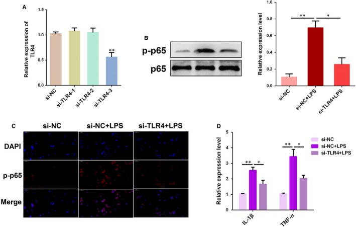
Figure 5.

Knockdown of TLR4 attenuates LPS‐induced inflammatory response. A, BEND cells were transfected with TLR4 siRNA (si‐TLR4) or the negative control (si‐NC) for 24 h and then stimulated with LPS for 12 h. The TLR4 expression was determined by qPCR. B, The protein level of NF‐κB p65 was measured by Western blotting. C, Translocation of the NF‐κB p65 subunit from the cytoplasm into the nucleus was assessed by immunofluorescence staining. Blue spots represent cell nuclei, and red spots indicate p‐p65 staining. D, The mRNA levels of pro‐inflammatory cytokines IL‐1β and TNF‐α were detected by qPCR. Data are presented as the mean ± SEM. *P < .05; **P < .01
