
Figure 3.
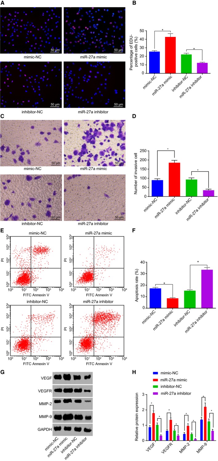
miR‐27a inhibition restrains PC cell proliferation and invasion but accelerates apoptosis. A and B, Positive cell proliferation in PC in response to the treatment of miR‐27a mimic or inhibitor, as detected by EdU (200×). C and D, Cell invasion in PC in response to the treatment of miR‐27a mimic or inhibitor (200×). E and F, Cell apoptosis in PC in response to the treatment of miR‐27a mimic or inhibitor. G and H, Protein levels of VEGF, VEGFR, MMP‐2 and MMP‐9 in response to the treatment of miR‐27a mimic or inhibitor. *P < .05 vs PANC‐1 cells treated with mimic‐NC or inhibitor‐NC. The above data are measurement data and described as mean ± standard deviation. Comparisons among multiple groups are analysed by one‐way analysis of variance. The experiment is repeated three times independently. miR‐27a, microRNA‐27a; PC, pancreatic cancer; NC, negative control; EdU, 5‐ethynyl‐2’‐deoxyuridine; VEGF, vascular endothelial growth factor; VEGFR, vascular endothelial growth factor receptor; MMP, matrix metallopeptidase