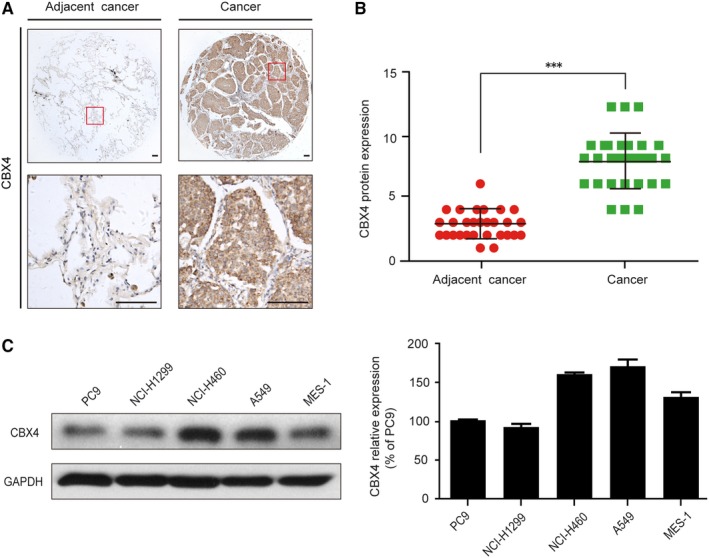
Figure 1.

The relative expression of CBX4 was elevated in human lung cancer tissues and cells. A, Representative immunohistochemical images of CBX4 in human lung cancer and adjacent cancer tissues. Scale bars: 100 μm. B, The CBX4 expression levels in lung cancer and adjacent cancer tissues (n = 30). C, The expression level of CBX4 was analysed by Western blot in lung cancer cells (PC9, NCI‐H1299, NCI‐H460, A549 and MES‐1 cells) and the histograms show the CBX4 relative expression. Densitometric measurements were analysed using Quantity One software. The protein expression levels were normalized to those of the vehicle control (100%). Data are presented as means ± SD (*** P < .001)
