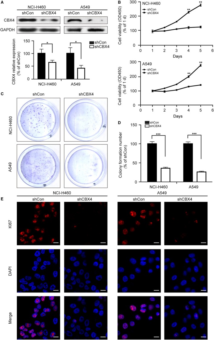
Figure 3.

CBX4 promotes the proliferation of lung cancer cells. NCI‐H460 and A549 cells were infected with shCon or shCBX4 lentivirus. A, The knockdown efficiency of shCBX4 was measured by Western blotting. B, Cell viability was measured by a microplate reader at 450 nm using CCK8 assay at the indicated time. (C and D) In colony formation assay, NCI‐H460 and A549 cells were fixed and stained with crystal violet after culturing for 2 wk, and the stained cells were counted in three independent experiments. E, NCI‐H460 and A549 cells were immunostained with Ki67 (red) and DAPI (blue). Photos were obtained by confocal microscopy and measured by Zeiss LSM Image Examiner software. Scale bars: 20 μm. All data were presented as mean ± SD (* P < .05, ** P < .01, *** P < .001)
