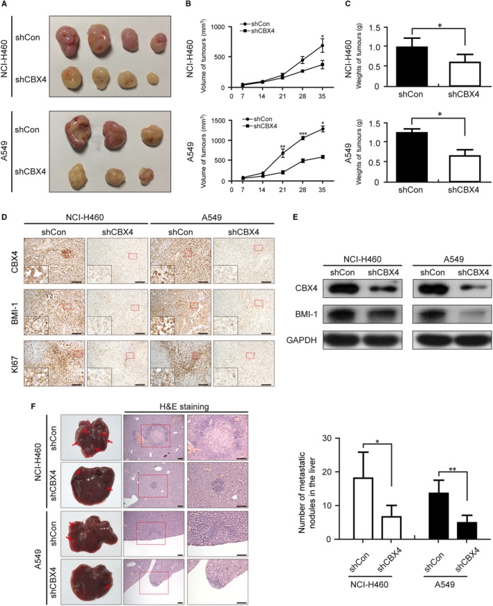
Figure 7.

CBX4 knockdown reduces the proliferation and metastasis of lung cancer in vivo. NCI‐H460 and A549 cells were injected into the corresponding part depending on the different models. A, After 5 wk, the nodular tumour was harvested and (B) the volumes of the tumour were measured every other week. The tumours in the CBX4‐silencing groups were obviously smaller than in the control group. C, The weights of the xenografts were measured at the end of experiment. D, Images for CBX4, BMI‐1 and Ki67 staining are shown for the xenografts. Scale bars: 100 μm. E, The expressions of CBX4 and BMI‐1 in xenograft tumours were analysed by Western blotting. F, Representative livers and H&E staining of the metastatic nodules in the livers were from the indicated groups. Metastatic nodules are pointed out by arrows. Statistical results of the metastatic nodules are presented as mean ± SD (* P < .05, ** P < .01, *** P < .001)
