FIGURE 1.
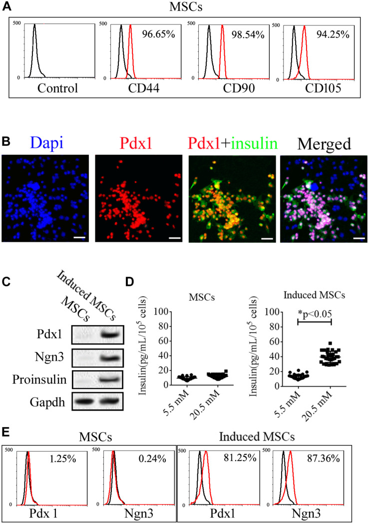
Expression of specific markers in uninduced and induced mice umbilical cord MSCs. (A) The isolated MSCs were positive for the MSC surface markers, CD44, CD90, and CD105, based on flow cytometry. (B) Mice umbilical cord MSCs were induced to differentiate into pancreatic beta cells with the use of a cocktail of factors, following which, the expression of the islet hormones, Pdx1 and insulin, was confirmed with immunofluorescence microscopy. (C) Based on western blotting, the induced MSCs exhibited expression of the islet hormones, Ngn3, Pdx1, and insulin, while the uninduced MSCs showed no expression. (D) Glucose-induced insulin secretion (GSIS) as measured by ELISA indicated that the insulin secretion of induced MSCs but not uninduced MSCs increased when treated with increasing glucose concentrations (n = 20; paired two-tailed t-test). (E) Expression of the islet markers, Pdx1 and Ngn3, in the induced MSCs was confirmed by flow cytometry.

