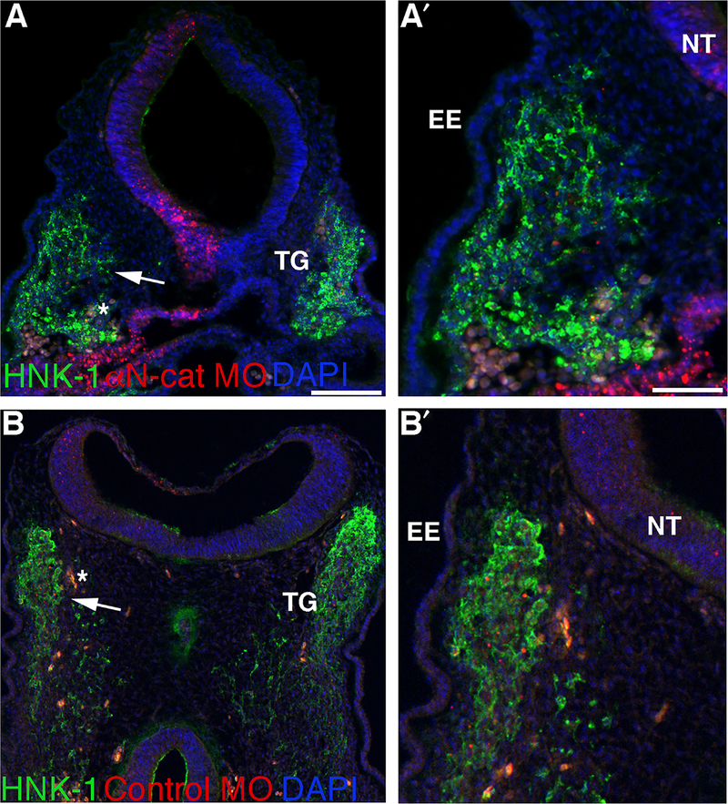
Fig. 3.
Morpholino-mediated depletion of αN-catenin increases the migratory neural crest cell contribution to the forming trigeminal ganglion in vivo. (A,B) Representative transverse sections taken at the axial level of the forming trigeminal ganglia of an HH15+ (A,A′) and HH15 (B,B′) embryo following αN-catenin (A) or control (B) MO (red) electroporation earlier in development and HNK-1 (green) immunohistochemistry after re-incubation. Arrows in (A,B) point to the region shown at higher magnification in (A′,B′). DAPI (blue) labels cell nuclei in all images. Asterisk (*) in (A,B) indicates blood cells. EE, embryonic ectoderm; NT, neural tube; TG, trigeminal ganglion. Scale bars in(A) and (A′) are 100 and 50 μm, respectively, and applicable to (B) and (B′).

