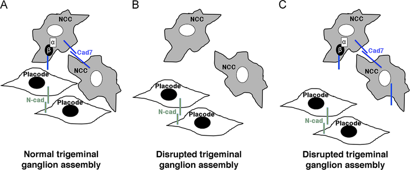
Fig. 9.
Model depicting the role of αN-catenin in neural crest cells during the assembly of the trigeminal ganglion. (A) Neural crest cells (NCC) and placode cells intermingle and coalesce to form the trigeminal ganglion. αN-catenin and Cadherin-7 (Cad7) are localized to neural crest cells, while placode cells express N-cadherin (N-cad). (B) Depletion of αN-catenin expands the migratory neural crest cell domain, but these neural crest cells possess reduced levels of Cad7. This prevents the formation of an effective scaffold for placode cells, leading to abnormalities in the formation of the trigeminal ganglion. (C) αN-catenin overexpression reduces the migratory neural crest cell domain, and these neural crest cells possess increased levels of Cadherin-7 on the surface of neural crest cells. The decrease in neural crest cells in the trigeminal ganglion-forming region may preclude effective interactions with placode cells. As in (B), the trigeminal ganglion is not assembled correctly. In (B) and (C), placode cell–placode cell interactions may be favored over placode cell–neural crest cell interactions, further enforcing the aberrant assembly of the trigeminal ganglion. α, αN-catenin; β, β-catenin.

