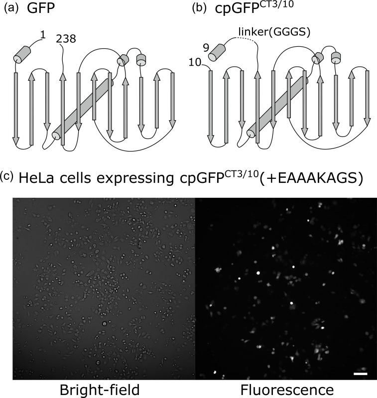
Fig. 1.
Circular permutation of GFP. (a) Schematic of GFP, including superfolder GFP. Rods indicate helices and arrows indicate β-sheets. (b) Schematic of cpGFPCT3/10. In (a) and (b), numbers indicate the first and last amino acids of superfolder GFP and cpGFPCT3/10, respectively. (c) Bright-field (left) and fluorescence (right) images of HeLa cells expressing cpGFPCT3/10(+EAAAKAGS). (Scale bar: 100 μm)

