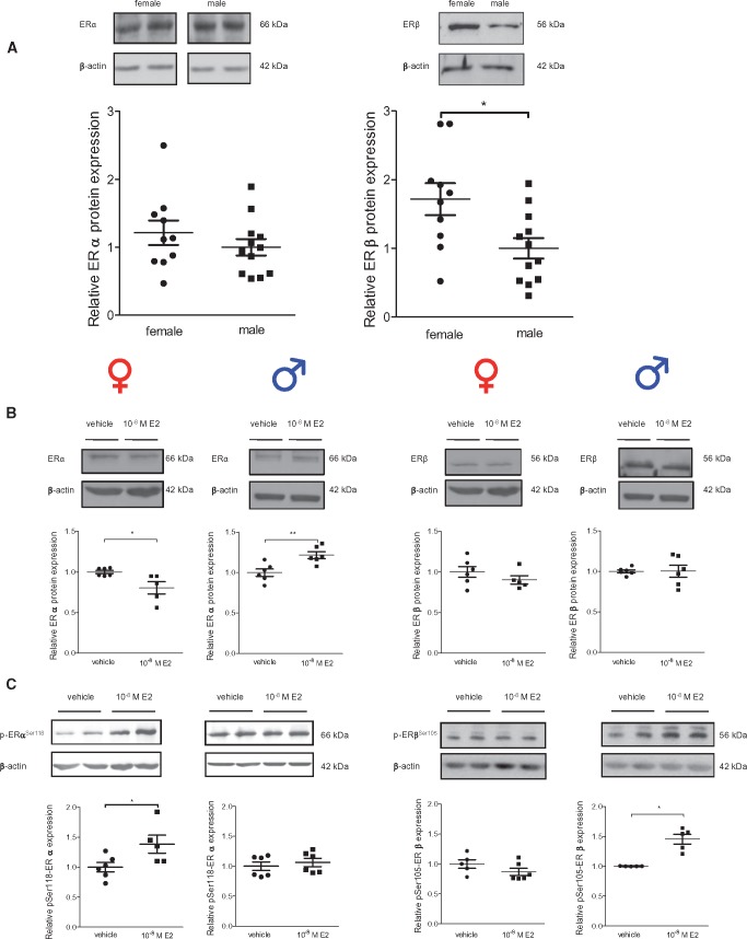
Figure 4.
ERα and ERβ protein and phosphorylation of ERα at serine 118 and ERβ at serine 105 differ between cardiac fibroblasts from male and female rats. (A) ERα and ERβ protein were investigated in non-treated female and male rCF. (B) ERα and ERβ protein was analysed in female and male rCF after 24 h of vehicle or 10−8 M E2-treatment. (C) Phosphorylation level of Ser118 of ERα and (D) of Ser105 of ERβ was analysed in female and male after 24 h of vehicle or 10−8 M E2-treatment. Probes were analysed by western blot and representative immunoblots of ERα ERβ, P-Ser118-ERα, P-Ser105-ERβ, and β-actin are shown. The expression of ER and P-ER were quantified and normalized to β-actin. In non-treated cells (A), relative protein levels of ERα and ERβ in female rCF are given as fold induction of male rCF. Protein and phosphorylation levels of ER of E2-treated cells (B–D) are given as fold induction of vehicle-treated cells. Mean ± SEM; ER comparison between sexes in non-treated rCF: female: n = 10 and male: n = 12; E2-treatment: from five to six independent samples derived from three independently repeated experiments; **P ≤ 0.01 and *P ≤ 0.05 comparison was performed using Mann–Whitney U-test.

