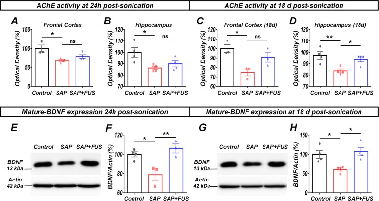
Fig. 3.
FUS increases AChE activity BDNF expression levels in a dementia rat model. a Twenty-four hours after sonication, AChE activity was significantly decreased in FC and b the hippocampus. c Eighteen days after sonication, FUS-mediated BBB opening induced a significant increase in AChE activity in FC and d the hippocampus. e Immunoblotting analysis shows BDNF protein expression levels in the hippocampus 24 h after sonication. BDNF levels in the FUS group increased significantly compared with those in the SAP and control groups. f Bar graph represents BDNF expression levels in the hippocampus. g Eighteen days after sonication, BDNF expression in the hippocampus in the FUS group increased significantly compared with that in the SAP and control groups. h Bar graph represents BDNF expression levels in the hippocampus. Data are expressed as mean ± SE. n = 3–4 for each group. *P < 0.05, **P < 0.01; one-way ANOVA with Tukey’s multiple comparisons test

