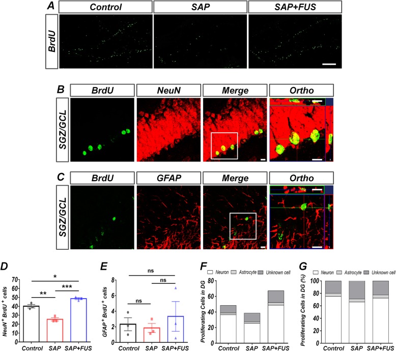
Fig. 5.
FUS increases neurogenesis and does not affect gliogenesis in a dementia rat model. a Representative photographs show the distribution of survived proliferation of BrdU-labeled positive cells in SGZ/GCL of DG of the hippocampus 18 days after sonication. The scale bar represents 200 μm. b Representative photographs of BrdU (green, a proliferative cell marker) and NeuN (red, a neuron marker) and c BrdU (green, a proliferative cell marker) and GFAP (red, an astrocyte marker) double-labeled cells in SGZ/GCL of DG of the hippocampus at 18 days after sonication. The scale bar represents 20 μm. d Quantification of BrdU and NeuN double-labeled cells. Compared with the SAP and control groups, the SAP+FUS group showed a significant increase in BrdU/NeuN-positive cells. e No significant differences were found in the numbers of BrdU/GFAP-positive cells among the groups. f Survived newly generating cells were determined, indicative of neurogenesis and gliogenesis. g The overall proportion of cells with the SGZ/GCL phenotype in DG of the hippocampus among the groups. Data are expressed as mean ± SE. n = 3–4 for each group. *P < 0.05, **P < 0.01; one-way ANOVA with Tukey’s multiple comparisons test

