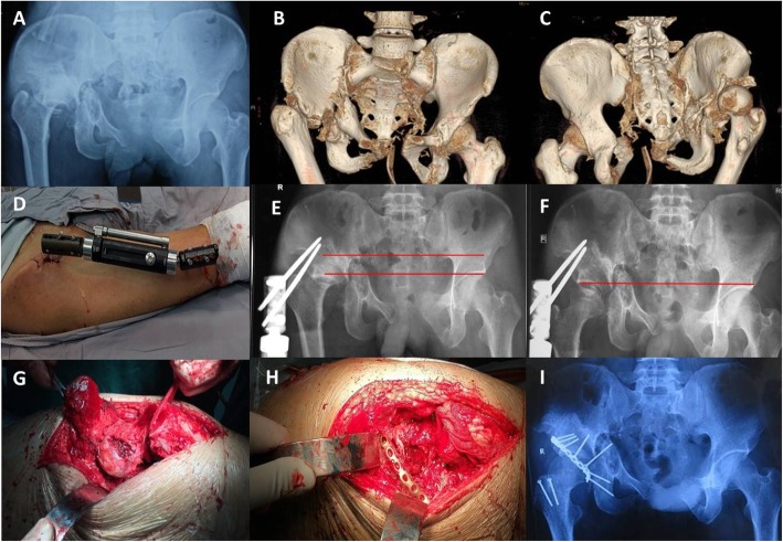
Fig. 1.
Patient 12. a-c Preoperative X-ray and three-dimensional reconstruction computed tomography images 6 months after injury. d-e External fixation placement. f The femoral head was drawn underneath the articular surface after 31 days of traction. g-h, k-l incision, open reduction with osteotomy of the greater trochanter, and fixation of posterior wall of acetabulum. i Postoperative X-ray image showed correction of limb length discrepancy and reduction of femoral head was originated from our previous study [16] and was authorized by Chinese Journal of Orthopaedic Trauma

