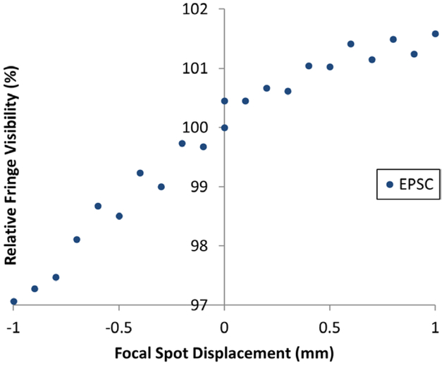
Figure 5.
Relative fringe visibility as a function of x-ray tube focal spot displacement. Normalized fringe visibility values at the imaging condition of 40 kVp/1.0 mA are plotted for cross-grating electromagnetic phase stepping. The changes were within a total range of 4.5% for a bidirectional spot displacement ±1.0 mm.

