Figure 2.

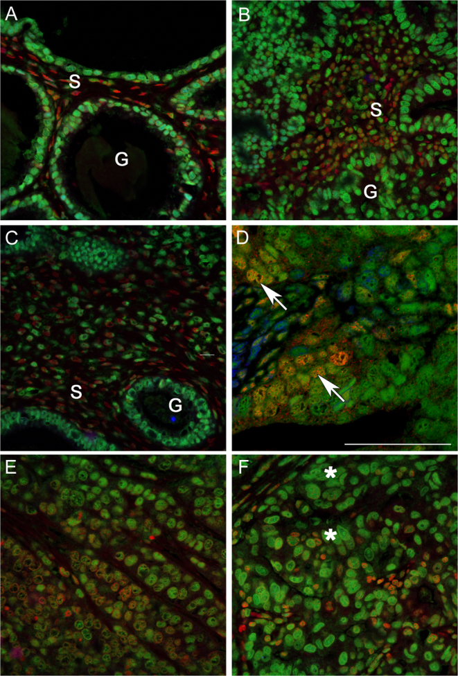
Co-localisation of ERα and ERβ5 in stage 1 endometrial adenocarcinomas identifies variable co-expression of both proteins in a subset of epithelial cells. Examples of staining in endometrial cancer tissues classified by a pathologist as G1 well (A and B), G2 moderately (C and D) or G3 poorly (E and F) differentiated. Note glands (G) surrounded by a single layer of epithelial cells could be identified in well and some moderately differentiated tissue associated with a stromal compartment (S) containing fibroblasts (s). The architecture of the poorly differentiated cancers was less organised and dominated by epithelial cells. Intense immunostaining for ERβ5 (green, asterisks) as well as evidence of co-expression of ERα (yellow-red, arrows) was detected in epithelial cells.

 This work is licensed under a