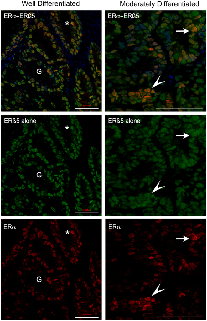
Figure 3.

Confocal imaging identifed epithelial cells in endometrial cancers with variable amounts of ERα and ERβ5 proteins. Confocal images typical of endometrial cancers classified as well or moderately differentiated are illustrated showing merged (top panel) and individual channels for ERβ5 (green, middle) and ERα (lower red). The intensity of immunostaining for ERβ5 appeared similar between different nuclei within each of these samples whereas the amount of protein in nuclei stained with an antibody specific for ERα (red) revealed a range of intensities from low to high with the latter identifed by yellow/orange staining in the merged image (examples * and arrowhead). Scale bars 50 µm.

 This work is licensed under a