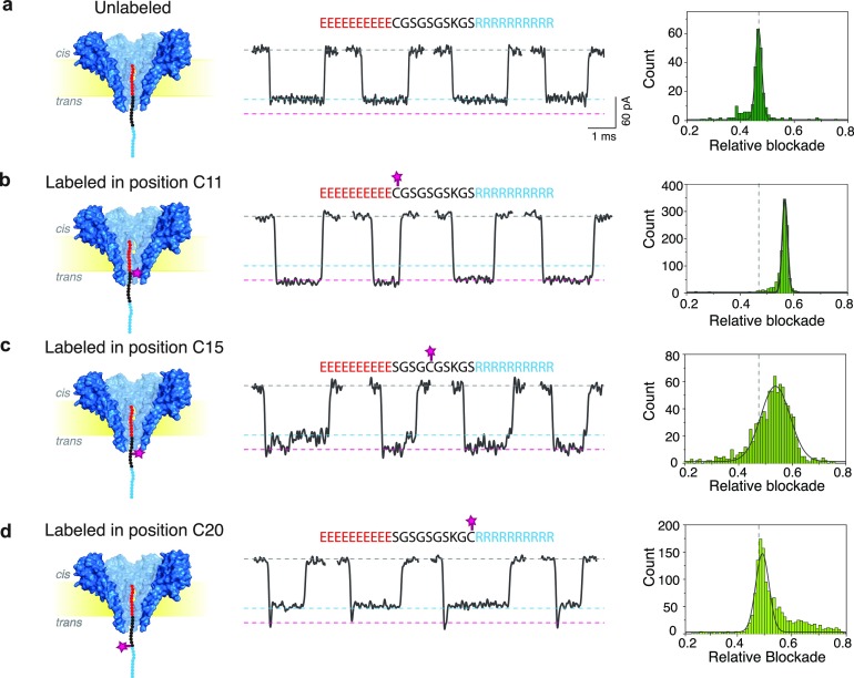
Figure 2.
Peptide labeled at different positions through FraC. From left to right: Schematic, current traces, and relative blockade histograms for unlabeled peptide (a) and for fluorescein-labeled peptide at positions C11 (b), C15 (c), and C20 (d). All labeled peptide samples were HPLC purified and verified using mass spectrometry. Measurements were done in a buffer containing 1 M NaCl, 10 mM Tris, and 1 mM EDTA at pH 7.5. Peptides were added to the cis compartment and measured at −90 mV.

