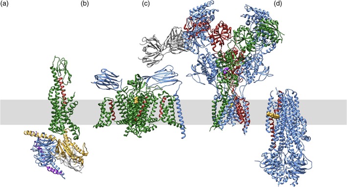
Figure 3.

Ribbon drawings of exemplar structures from each of the four classes of membrane‐bound proteins of pharmacologic interest, viewed parallel to the lipid bilayer (shaded grey rectangle). (a) GPCR (PDB 5vai)36: GLP1‐R (glucagon‐like peptide‐1 receptor, active conformation in green) bound to GLP1 (red) and heterotrimeric G‐protein (blue, yellow, and magenta). (b) VGIC (PDB 6j8i)106: Nav1.7 (green), beta1 and beta2 (blue), bound to inhibitor tetrodotoxin (yellow). Voltage‐sensing helices are shown in red. (c) LGIC (PDB 5uow)72: NMDA receptor (blue, green, and red) bound to channel blocker MK‐801 (magenta). An antibody Fab (grey) was used in the structure determination. (d) Transporter (PDB 6o2p)85: CFTR (cystic fibrosis transmembrane conductance regulator (blue) bound to ivacaftor (yellow), which interacts with a long transmembrane helix involved in gating (red)
