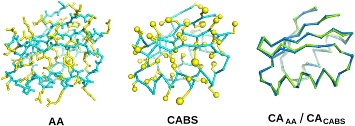
Figure 1.

Representation of the three‐dimensional structure of the α + β type 2‐layer sandwich protein (PDB ID: 3IWL). The first panel shows all‐atom (AA) structure. The second panel shows a model in the CABS representation. For both cases, side‐chain atoms are shown in yellow (for CABS—the C‐beta atom and the united side‐chain pseudo‐atom) while the backbone is shown in cyan. The third panel shows the superposition of the alpha carbons from the CABS model (blue) and the experimental structure (green)
