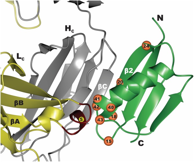
Figure 2.

Interface between Protein G (green) and Fab (yellow–gray). Residues that were diversified in the phage display mutagenesis library to convert Protein G to GA1 are numbered and marked as orange spheres. Region in the Lc of FabS that was diversified to generate the affinity matured FabLRT involved residues 123–127 marked in red. The position of E123S mutation that differentiates between the FabH and FabS scaffolds is marked by a yellow sphere
