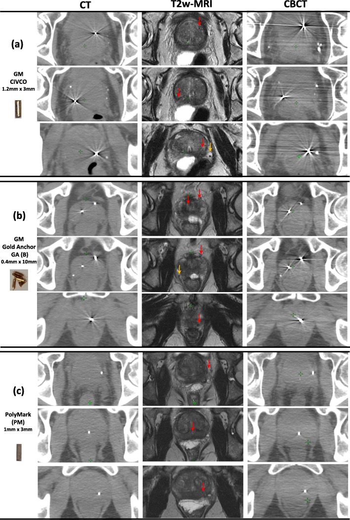
Fig. 4.
Prostate gland of three FM on three different patients as they appear on axial slices of CT/CBCT and T2w-MRI. Each patient had 3 fiducial markers. Patient 1 (a) GM CIVCO; routinely used in our institute for IGRT, patient 2 had GM GA (implanted as a ball shape) (b) and patient 3 had a PM polymer markers (c). FM indicated with red arrows on MRI and the orange arrows point to natural calcifications

