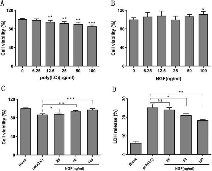
Fig. 1.
NGF protected cells against TLR3-induced cytotoxicity. a-b Cell viability was determined by a CCK-8 assay in HCECs cultured with various concentrations of poly (I:C) or NGF for 24 h. c Cell viability was determined by a CCK-8 assay in HCECs co-cultured with NGF (range from 25 ng/ml to 100 ng/ml) and poly (I:C) (25 μg/ml) for 24 h. NGF significantly increased cell viability. d Cytotoxicity was determined by a LDH assay when HCECs were co-cultured with NGF (range from 25 ng/ml to 100 ng/ml) and poly (I:C) (25 μg/ml) for 24 h. NGF significantly suppressed LDH release in HCECs. LDH release was presented as percentage of control (whole cell lysis, 100%). Significant differences (**p < 0.01, ***p < 0.001) existed compared to blank group (without poly (I:C) or NGF), conducted by one-way ANOVA and Dunnett-t test. Significant differences existed between poly (I:C) and poly (I:C) + NGF treatment group, conducted by two-tailed student’s t test. Data represented mean ± SD from six independent representative experiments in each condition

