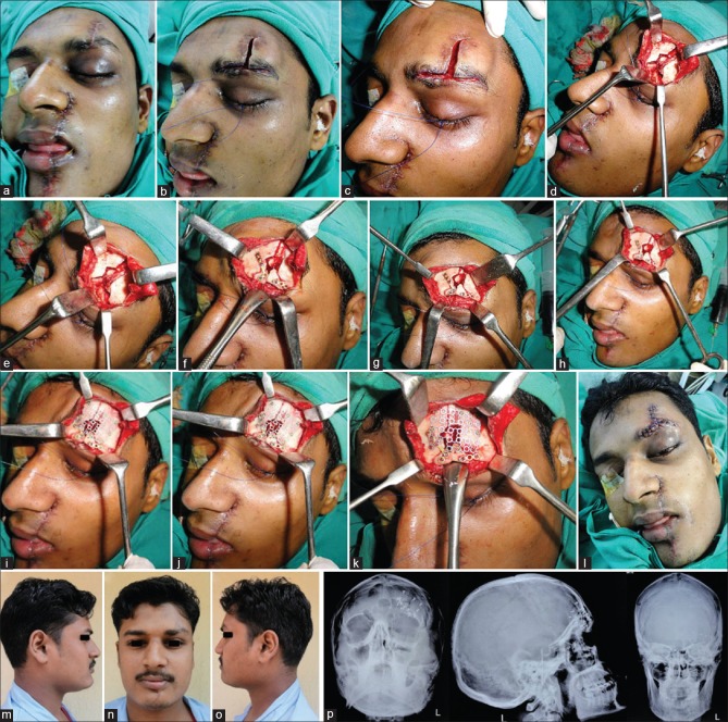
Figure 9.
(a-d) Existing scar used to expose fracture of the frontal bone and left supraorbital rim. (e-h) Displaced fragments reapproximated, repositioned, and fixed. Unsalvageable free fragments of crushed bone removed. (i-l) Defect of the frontal bone reconstructed using three-dimensional dynamic titanium mesh implant, followed by layer-wise closure. (m-o) Smooth postoperative recovery with a good esthetic as well as functional outcome. (p) Postoperative radiographs showing restoration of the displaced frontal bone and orbital roof and rim fractures with implants in situ

