Figure 3.

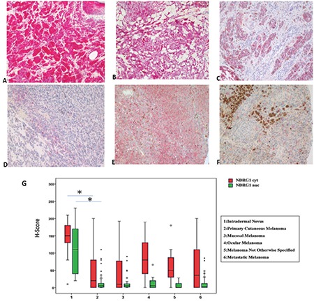
a-g. NDRG-1 immunostaining in study groups. Strong positivity is easily detected in intradermal nevus (a), weak and heterogenous positivity is detected in primary cutaneous (c), rectal (d), and metastatic melanoma (f). However, some primary melanomas (b) and ocular melanomas (e) show more intense positivity, though not as strong as in intradermal nevus. Nuclear and cytoplasmic scores of NDRG-1 in nevus are higher than melanomas (g). Original magnifications a, b, c ×200, d, e, f, ×100. Box-plot graphs for NDRG-1 immunostaining H-scores of study groups (g).
NDRG-1: N-myc downstream regulated gene-1
