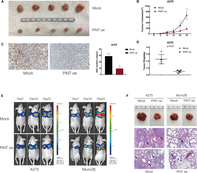
FIGURE 4.
LINC-PINT inhibited melanoma progression in vivo. (A) Mock and PINT oe A375 cells were injected into the nude mice. Tumors derived from cells with (lower panel) or without (upper panel) LINC-PINT overexpression were removed from the mice. (B) Tumor volume was evaluated every 5 days after the injection of Mock (n = 5) and PINT oe (n = 5) A375 cells for 25 consecutive days. ∗p < 0.05, ∗∗p < 0.01. (C) Ki-67 staining of Mock and PINT oe tumor tissues. (D) The weights of Mock (n = 5) and PINT oe (n = 5) A375 tumors. (E) Effect of LINC-PINT on tumor metastasis in a lung metastasis mouse model. Mock and PINT oe A375 and Mum2B cells were injected into the caudal vein of nude mice (n = 3 for each group). (F) Representative lung tissues and their HE-stained sections (100× magnification) are shown. The black arrows showed the metastases.

