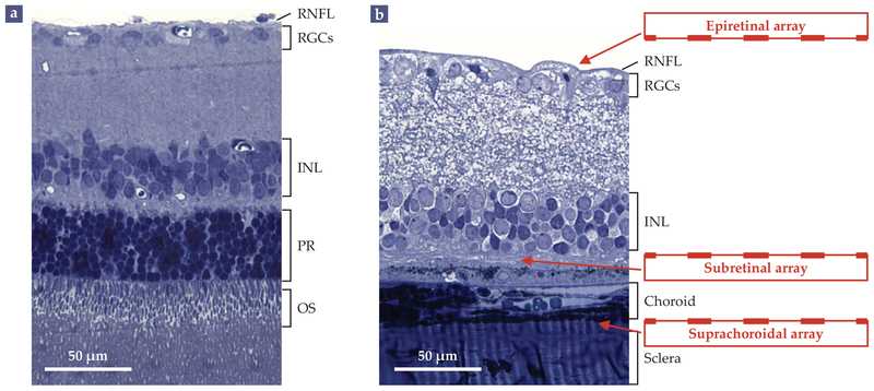
FIGURE 2. RETINAL TISSUE, FOR BETTER AND WORSE.
(a) A histological cross section shows the layers of a healthy rat retina. Absorption of light (incident from the top) by the outer segments (OS) of photoreceptor cells (PR) at the back of the retina generates electrical and chemical signals that are processed by the neurons of the inner nuclear layer (INL) and by the retinal ganglion cells (RGCs). The RGCs generate spike-like signals known as action potentials that propagate via the retinal nerve-fiber layer (RNFL) to the brain. (b) In a rat with retinal degeneration, the photoreceptor cells have all but wasted away. Vision can be partially restored by introducing electrode arrays on top of the nerve-fiber layer (epiretinal array), below the inner nuclear layer (subretinal array), or between the underlying choroid and sclera layers (suprachoroidal array). (Adapted from G. A. Goetz, D. V. Palanker, Rep. Prog. Phys. 79, 096701, 2016.)

