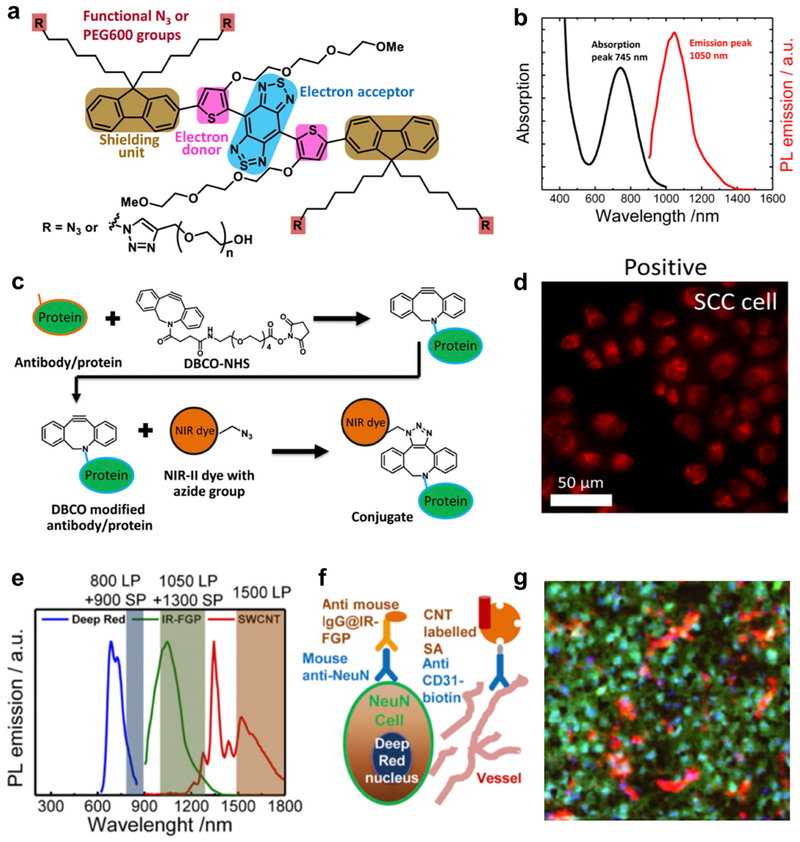
Figure 1. NIR-II fluorophore IR-FGP with high QY for the molecular imaging application.
a) Molecular structure of NIR-II fluorophore IR-FGP. The fluorophore is modified with active functional groups -N3 for subsequent bio-conjugation. b) Emission and adsorption spectra of IR-FGP. c) Scheme of conjugation of IR-FGP with proteins under mild copper-free click chemistry. d) In vitro staining of EGFR positive SCC cell line using conjugate Erb@FGP. e) PL emission spectra of Deep Red, IR-FGP, and NIR-IIb SWCNT including the long-pass/short- pass filter ranges used for three-color imaging. Three ranges without overlapping were chosen for the multi-color molecular imaging application. f) Scheme regarding how three-color molecular imaging is performed. Deep Red was used to stain nucleus. IR-FGP was chosen to stain the neuron cells. CNT was applied to stain vessels. g) Multicolor brain tissue imaging with high resolution by NIR-II confocal microscopy. Reproduced with permission[6c]. Copyright 2017, National Academy of Sciences.

