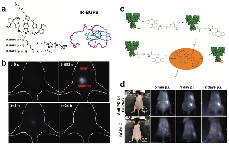
Figure 4. Molecular imaging of immune checkpoint PD-L1 based on the renal-excreted NIR-II fluorophores.
a) Molecular structure and molecular dynamics simulation of renal-excreted NIR-II fluorophore IR-BGP6. b) Renal excretion behavior of IR-BGP6. Most IR-BGP6 went to bladder after intravenous injection. c) Scheme of conjugation of PD-L1 mAb onto IR-BGP6. d) Non-invasive molecular imaging of PD-L1 using anti-PD-L1-BGP6 probe. Without attachment of PD-L1 mAb negligible IR-BGP6 accumulated within the MC38 tumors. Reproduced with permission[6b]. Copyright 2018, John Wiley and Sons.

