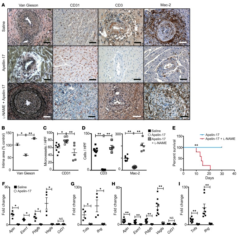
Figure 6. Apelin-17 analog treatment suppresses arterial vasculopathy and immune cell infiltration of heart transplants.
Apln+/y male hearts were transplanted into WT female recipients. Two weeks after transplantation the recipient mice were treated daily with saline or apelin-17 analog or with apelin-17 analog plus l-NAME; then the heart allografts were recovered at the time the graft heartbeat stopped, or at 6 weeks after transplantation. (A) Photomicrographs of transplanted hearts stained with van Gieson, immunostained for CD3, Mac-2, or EC CD31. Scale bars: 50 μm. (B) Quantitation as in Figure 2 of the intima area of graft arteries in heart recipients treated with saline (n = 8 biological replicates), apelin-17 analog (n = 9), or apelin-17 analog plus l-NAME (n = 5). (C) Quantitation of CD31+ microvessels in grafts of heart recipients treated with saline (n = 8 biological replicates), apelin-17 analog (n = 9), or apelin-17 analog plus l-NAME (n = 5). (D) Quantitation of immune cell infiltration in myocardium of grafts from heart recipients as in C. Mean ± SEM; *P < 0.05, **P < 0.01 by 1-way ANOVA with Bonferroni’s post hoc test. (E) Survival of heart allografts in mice treated with apelin-17 analog without (n = 9) or with l-NAME (n = 5 biological replicates) starting on day 14 after transplantation. **P < 0.01 by log rank. (F and G) Expression of endothelial repair (F) and proinflammatory (G) genes in microdissected coronary arteries from recipient mice treated with saline (n = 4 pairs) or apelin-17 analog (n = 5 pairs). Coronary artery data were analyzed by Mann-Whitney test. Mean ± SEM; *P < 0.05, **P < 0.02. (H and I) Expression of endothelial repair (H) and proinflammatory (I) genes in myocardium of heart grafts from recipient mice treated with saline (n = 8) or apelin-17 analog (n = 9). Mean ± SEM; **P < 0.01 by Student’s t test.

