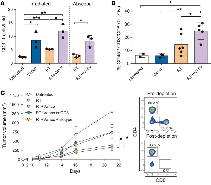
Figure 2. The effects of vancomycin are abrogated in CD8-depleted mice.
(A) Quantification of CD3+ cell infiltration by immunohistochemistry of B16-OVA–derived primary tumor sections from untreated, vancomycin treatment alone (Vanco), RT treatment alone (RT), or vancomycin plus RT combination treatment (RT+Vanco), and of abscopal tumor sections from RT abscopal or RT+Vanco abscopal combination treated. Mean ± SEM are shown. Statistical significance was assessed by Tukey’s test. (B) Primary tumors from each treatment group were digested and individual cells were analyzed by flow cytometry to determine the percentage of OVA-specific CD8+ T cells (CD45+/CD3+/CD8+/TET-OVA). Mean ± SEM are shown. Statistical significance was assessed by Tukey’s test. (C) Tumor growth rates in CD8-depleted mice. n = 5 to 10 mice per group. Mean ± SEM are shown. Statistical significance was assessed by 2-way ANOVA. Data are representative of at least 2 independent experiments. *P < 0.05, **P < 0.001, ***P < 0.01.

