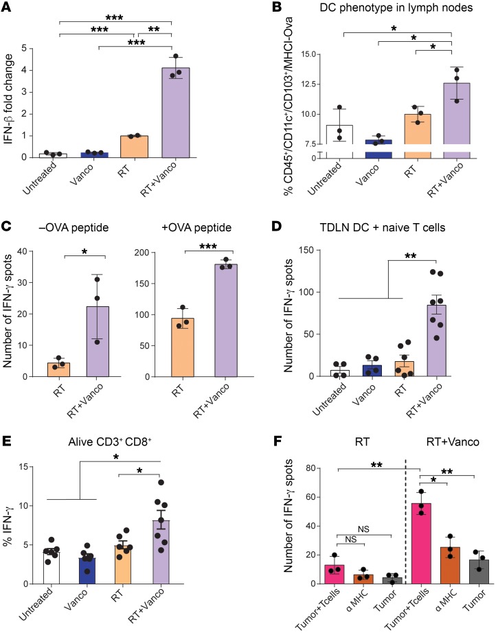
Figure 4. Vancomycin treatment increases local TAA cross-presentation and antigen recognition in tumors.
(A) Ifnb1 mRNA expression levels in TDLNs 1 day after irradiation. (B) Anti-MHC1 (Kb)-SL8 OVA peptide staining on tumor-infiltrating CD11c+ CD103+ DCs 5 days after RT. (C) IFN-γ ELISPOT assay plated with TDLN single-cell suspension and OT1 T cells (1:5 TDLN cells/T cells) in absence (left) or presence (right) of OVA peptide. Cells were harvested 5 days after RT treatment. (D) Coculture of purified CD11c+ DCs from TDLNs from each treatment group were incubated overnight with naive OT1 T cells in an IFN-γ ELISPOT assay (1:10 DCs/T cells). (E) Percentage of IFN-γ expression from overnight OT1 cells cocultured with DCs from mice treated with each therapeutic approach. (F) B16-OVA tumors from mice treated with RT alone or with the RT and vancomycin (RT+VANCO) combination treatment were dissociated and plated with OT1 cells in an IFN-γ ELISPOT plate for 24 hours. n = 5 to 10 mice per group. Data are representative of at least 2 independent experiments. Mean ± SEM are shown. Statistical significance was assessed by Tukey’s test. *P < 0.05, **P < 0.01, ***P < 0.001.

