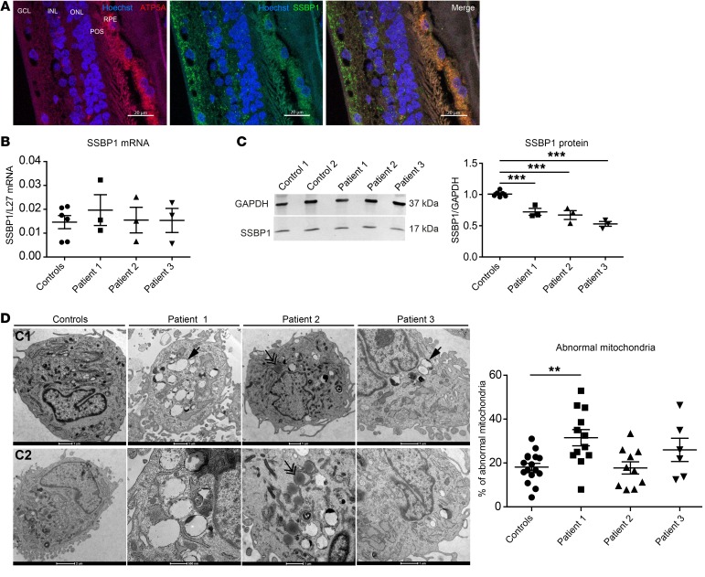
Figure 3. SSBP1 protein expression in human retina and fibroblasts.
(A) SSBP1 protein expression in human retina. Immunofluorescence labeling was done on retinal cross-sections from healthy human donor. Hoechst was added to label nuclei (blue). SSBP1 localization was done using a specific antibody (green). An antibody against the ATP synthase subunit 5A was used to target mitochondria (red). RPE, retinal pigment epithelium; POS, photoreceptor outer segment; ONL, outer nuclear layer; INL, inner nuclear layer; GCL, ganglion cell layer. Scale bars: 20 μm. (B) Quantification of SSBP1 transcript levels in cultured skin fibroblasts from both controls and affected individuals (patient 1, patient 2, and patient 3). mRNA levels were normalized to the reference gene L27. (C) Western blot in lysates from controls (control 1 and control 2) and patient fibroblasts and densitometric analysis of SSBP1 protein abundance in lysates from both controls and patient fibroblasts. GAPDH was used as a loading control. Data are shown as mean ± SEM. ***P < 0.001. (D) Representative ultrastructure of the mitochondria from both controls (C1, C2) and patient fibroblasts by TEM. Scale bar: 1 μm. Single arrows show abnormal mitochondria. Double arrows show lipid droplets. Quantification analyses of abnormal mitochondria (large vacuoles, disturbed cristae) in fibroblasts from both controls and patients. Data are represented as mean percentage of abnormal mitochondria ± SEM in total examined mitochondria. **P < 0.01. All data are representative of 3 independent experiments. One-way ANOVA with Dunnett’s correction was used.

