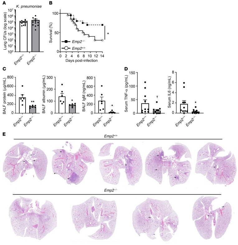
Figure 3. EMP2-null mice have reduced mortality and lung injury during bacterial pneumonia.
(A) Emp2+/+ and Emp2–/– mice were infected with K. pneumoniae by oropharyngeal aspiration and then had bacterial CFUs quantified in lung homogenates 24 hours after infection (n = 11–12/genotype). (B) Survival was monitored in mice infected with K. pneumoniae as in A (n = 20/genotype). (C) BALF protein, albumin, and IgM were measured in mice 48 hours after K. pneumoniae inhalation (n = 5–6/genotype). (D) Serum TNF-α and IL-6 were quantified by ELISA 48 hours after lung infection with K. pneumoniae. (E) Representative images of lungs from mice (n = 4–5/genotype; ×1 magnification) 48 hours after infection with K. pneumoniae by oropharyngeal aspiration. Inflammatory areas are indicated by arrows; arrowhead points to inflammation on the pleural surface. Inflammation (predominantly neutrophilic infiltration) was more severe in Emp2+/+ mice. Data in A, C, and D are mean ± SEM and are representative of at least 3 independent experiments. †P = 0.06; *P < 0.05; **P < 0.01 by unpaired 2-tailed Student’s t test or log-rank test (survival).

