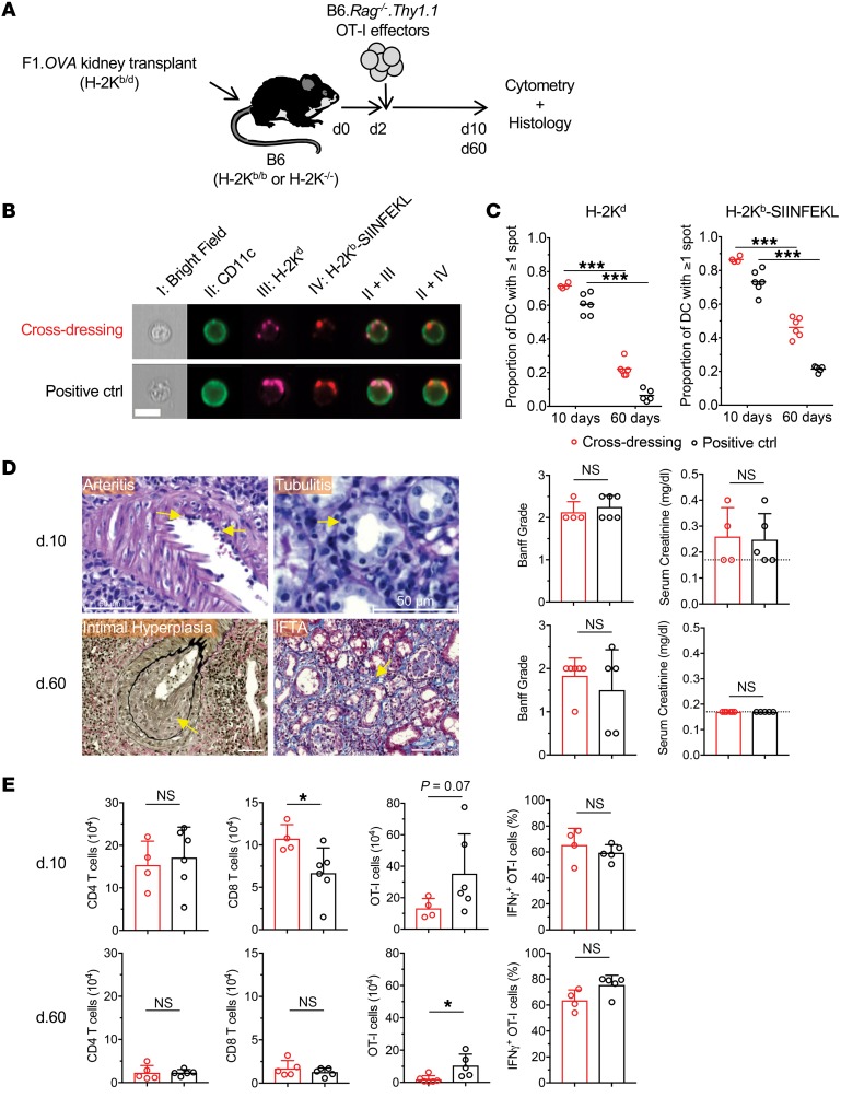
Figure 4. Cross-dressing of host DCs with donor MHC class I–peptide complexes in kidney allografts.
(A) (B6×BALB/c)F1.Act-mOVA (F1.OVA) (H-2Kb/d) kidneys were transplanted to B6 H-2Kb–sufficient (H-2Kb/b) WT (n = 5–6) or B6 H-2Kb-deficient (H-2K–/–) (n = 4–6) recipients. 1 × 10⁷ B6.Rag–/– OT-I CD8+ effector T cells, which recognize the OVA peptide SIINFEKL bound to H-2Kb, were transferred i.v. 2 days later. Grafts were analyzed 8 or 58 days after OT-I transfer. Control and experimental groups are shown in Table 2. (B) Leukocytes were isolated from transplanted allografts and analyzed by ImageStream. Intact H-2Kb–SIINFEKL complexes and donor H-2Kd molecules were identified as discreet spots on surface of host (CD11c+) DCs. Representative images from each group from day 10 grafts shown. On average, 426 (range = 153–784) and 3068 (range = 547–10366) cells were analyzed per animal on day 10 and day 60, respectively. (C) Proportion of host DCs positive for 1 or more spots of either H-2Kd or H-2Kb–SIINFEKL. Each data point represents analysis of 1 transplanted animal. (D) Histological analysis of grafts from cross-dressing and control groups removed on day 10 and day 60. Photomicrographs show examples of arteritis, tubulitis, intimal hyperplasia, and interstitial fibrosis and tubular atrophy (IFTA) in grafts from cross-dressed group. Scale bars: 50 μm. Bar graphs depict quantitation of Banff grades and serum creatinine on day 10 and day 60. Dashed line denotes lower detection limit for creatinine (<0.2 mg/dl). (E) Flow cytometric analysis of cellular infiltrate in allografts removed on day 10 and day 60 from cross-dressing and positive control groups. *P < 0.05; ***P < 0.001, 2-tailed unpaired t test.

