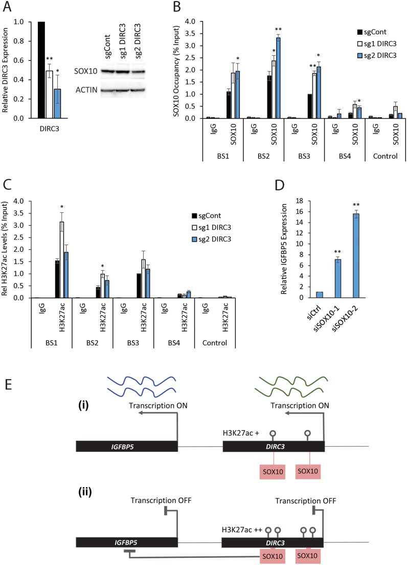
Fig 6. DIRC3 induces closed chromatin at its site of expression thereby blocking SOX10 DNA binding and activating IGFBP5.
ChIP assays were performed in DIRC3 depleted SK-MEL-28 and control cell lines using the indicated antibodies against either SOX10, H3K27ac or an isotype specific IgG control. (A) DIRC3 depletion was confirmed using qRT-PCR. Western blotting showed that SOX10 protein levels do not change upon DIRC3 knockdown. ACTIN was used as a loading control. (B) The indicated SOX10 binding sites were analysed by qPCR. % input was calculated as 100*2^(Ct Input-Ct IP). (C) DIRC3 depletion leads to an increase in H3K27ac levels at SOX10 bound regulatory elements within the DIRC3 locus. (D) SOX10 represses IGFBP5 expression. SOX10 was reduced in SK-MEL-28 cells using transfection of two independent siRNAs (see Fig 2C for SOX10 levels). IGFBP5 expression was quantified using RT-qPCR three days later. POLII was used as a reference gene and expression changes are shown relative to a non-targeting control (set at 1). (E) Model illustrating that DIRC3 acts locally to close chromatin and prevent SOX10 chromatin binding at melanoma regulatory elements within its locus. This leads to a block in SOX10 mediated repression of IGFBP5 and subsequent increase in IGFBP5 expression. All qPCR results are presented as mean values +/- SEM, n = 3. One-tailed student’s t-test p < 0.05 * p < 0.01 **.

