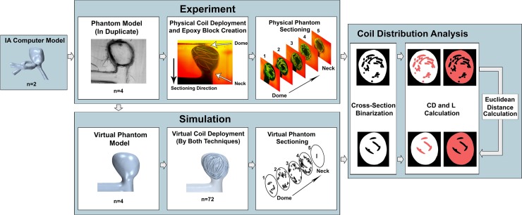
Fig 3. Flowchart of our experimental approach.
From Left to Right, computer models of 2 IAs were used in this study as testbeds. Experiment: Based on the IA models, 4 silicone phantoms (duplicates of each IA) were fabricated, coils were deployed into each phantom, and intra-aneurysmal cross-sections were extracted. Simulation: The phantoms were imaged to create virtual models, and coil deployments were virtually recreated using the Original Technique and Improved Technique (9 iterations for each phantom). Cross-sections were extracted matching those in the experiments. Coil Distribution Analysis: Physical and virtual coil distributions were quantified by coil density (CD, space occupied by coil–in red in left image) and lacunarity (L, gaps between coils–red in right image) from cross-section images. To compare the virtual techniques against experiments, Euclidean distances from the experimental coil distributions to the virtual coil distributions were calculated and evaluated (Coil Distribution Analysis).

