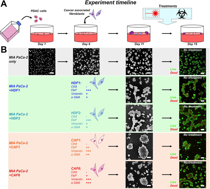
Fig. 1. Experiment timeline of the 3D co-culture models.
(A) Adherent 3D cultures were initiated on day 1 by seeding 7.5 × 10³ PDAC cells (MP2 cell line) on solidified Matrigel in each well of a 24-wells plate. Fibroblasts, either HDFs or CAFs, were added on day 8 at a density of 5 × 10⁴, corresponding to a 2:1 cancer cell:fibroblast ratio [11]. Cultures were maintained until day 11, after which treatments were initiated. Experiments were terminated on day 15, after which live/dead staining and image analyses were performed. (B) Cancer microtumor development in absence and presence of the various fibroblasts. In absence of fibroblasts, the PDAC cultures develop into relatively homogeneously dispersed adherent spheroids. In presence of HDFs, large interconnected networks of microtumors are formed. In presence of CAFs, multiple individual cancer spheroids are condensed into large microtumor masses.

