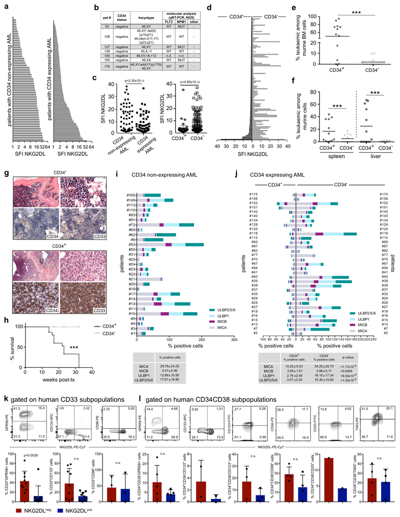
Extended Data Fig. 7. Surface expression of NKG2DLs in non-CD34-expressing versus CD34-expressing AML, analysis of in vivo leukaemogenicity in CD34+ versus CD34− subpopulations of AML cells and co-staining for NKG2DL and LSC markers.
a, Comparison of expression of NKG2DLs (SFI, specific fluorescence intensity indices) as measured by NKG2D–Fc in non-CD34-expressing (n = 57, left) and CD34-expressing (n = 107, right) cases of AML. b, Characteristics of patients with >99% NKG2DL+ cells among AML cells within our cohort (comprising n = 175 cases of AML; note that all cases are non-CD34-expressing cases of AMLs. c, Quantification of individual patient data shown in a, and of expression of NKG2DLs in CD34+versus CD34− subpopulations within CD34-expressing cases of AML for which individual data are shown in d. A two-sided Mann–Whitney test was used for statistical analysis. e–h, Analysis of sorted CD34+ and CD34− subpopulations of AML cells in in vivo xenotransplantation assays in NSG mice (no. 7, 8 and 12; n = 5 mice per AML and subpopulation). e, f, Flow cytometric analysis of summarized percentages of human leukaemic among mouse bone marrow (e), and spleen and liver cells (f). g, Histopathological bone marrow analysis using antibodies that recognize human, but not mouse, CD33 and CD34; 630× magnification. h, Kaplan–Meier analysis indicating mouse survival. A Mann–Whitney U test was used for statistical analysis. Note that CD34+ subpopulations with lower percentages of NKG2DL+ cells have increased stemness properties. i, j, Analysis of the percentage of positive cells with MICA, MICB, ULBP1 or ULBP2, ULBP5 or ULBP6 surface expression in non-CD34-expressing cases of AML (n = 29) (i) and in CD34+ and CD34− subpopulations of CD34-expressing cases of AML (n = 33) (j). In some cases the sum of the percentage of positive cells exceeds 100%, which reflects the fact that individual AML cells can express more than one NKG2DL. A two-sided Mann–Whitney U test was used for statistical analysis. k, l, Co-staining of NKG2DLs and LSC markers in CD33+ AML cells (k, n = 9 cases of AML, of which 9 out of 9 cases expressed GPR56 and CD133, and only 3 out of 9 cases expressed CD96) and within CD34+CD38− subpopulations of AML cells (l, n = 5 cases of AML; notably, in the other 7 cases of AML we analysed, all CD34+CD38− cells were found to lack expression of NKG2DLs). A two-sided Student’s t-test was used in k, l. Centre values represent mean, error bars represent s.d.

